It is a relatively safe procedure, complications like inflammatory reaction, pain, red eye, conjunctival scarring, extremely low eye pressure and further vision loss may occur in rare cases.
Glaucoma is one of the leading causes of irreversible blindness, with an estimated 12 million people affected in India. Among these, a staggering 1.2 million are blind due to this disease. It is aptly known as the “silent thief of sight” as patients commonly have no symptoms in the initial stages. Only as the disease progresses do they complain of visual problems. This leads patients affected by glaucoma to present late in their disease course. Treatment options are aimed at preserving existing vision and preventing further loss of vision.


One of the most commonly asked questions by patients is, “Is glaucoma curable?” Unfortunately, glaucoma is not curable and causes irreversible blindness. This, therefore, highlights the importance of early detection and treatment to preserve useful vision. Glaucoma is a lifelong disease that requires regular monitoring and lifelong follow-up.
It has consistently been voted the number #1 eye hospital in Karnataka since over a decade. It is a referral centre for difficult and complicated cases. We use a multidisciplinary approach to combat the challenges faced during the management of these cases. We have with us an impressive list of highly qualified and dedicated eye specialists who have specialized in caring for all forms of glaucoma
The glaucoma department saw over 1 lakh patients last year and has performed 1800+ glaucoma surgeries. These patients stand as testimony and brand ambassadors to the quality of care offered at NN.
Primary open angle glaucoma – is the most common type of glaucoma. In this type of glaucoma, the part of the eye through which the fluid of the eye flows out is open, permitting the outflow of fluid. However the drainage pathway may be defective leading to increased resistance to the outflow of fluid within the eye. Increased pressure may also occur due to increased production of fluid in a small percentage of cases. This type of glaucoma develops slowly without any symptoms. Initially it affects the peripheral or side vision and very gradually progresses to the centre. This is the reason why many people are not aware that they have the condition until they have significant vision loss affecting central vision.
Risk factors for primary open angle glaucoma are:
Angle closure glaucoma or closed angle glaucoma –is the second most common type of glaucoma which occurs due to narrow drainage channels in the eye. Gradual closing of the angle is called chronic angle closure and if the drainage angle closes suddenly, it causes an acute angle closure attack. Acute angle closure glaucoma usually presents as an emergency. A patient who is in an acute angle closure attack will have symptoms of eye pain, nausea, vomiting, redness, blurred vision and coloured halos due to a rapid increase in the eye pressure. In such cases the patient needs immediate treatment by an eye specialist.
Risk factors for angle closure glaucoma:
Normal tension glaucoma /Low tension glaucoma –In this type of glaucoma the optic nerve can get affected even though the pressure in the eye is normal. Although its cause is not entirely known or understood, normal tension glaucoma is believed to occur either because of an extremely fragile optic nerve that can get damaged even though the pressure in the eye is normal, or because of reduced blood flow to the optic nerve. Because of its silent nature, people usually do not have any visual complaints until a very advanced stage of the disease.
Risk factors for normal tension glaucoma are:
Glaucoma in infancy and childhood constitutes a rare but sight threatening group of diseases. Infants and young children with glaucoma typically present for ophthalmologic evaluation because the paediatrician or parents have noticed something unusual about the eyes. This is usually whitish discolouration and enlargement of the eyes, excessive watering, tendency to keep the eyes closed and sensitivity to light. Glaucoma in children can occur if a family member has glaucoma, the parents have had a consanguineous marriage or abnormal development during pregnancy.
This glaucoma is caused by abnormal intraocular fluid drainage from the eye as a result of a blocked or defective trabecular meshwork (the mesh-like drainage canals in the eye).In other cases, an abnormal drainage system may be the result of some other disease in the eye/ body which results in secondary glaucoma.
Congenital glaucoma is detected through a full eye examination which is usually carried out under sedation in the operation theatre, in case of babies and children under 3 years of age.Treating congenital glaucoma in its early stages can slow the development of the disease. In an uncomplicated case of congenital glaucoma, microsurgery can often correct the structural defects. Other cases are treated with medication and surgery.
There are certain other types of glaucoma where there is an identifiable cause for increased eye pressure resulting in optic nerve damage and vision loss. These are called secondary glaucoma. It may be caused by prolonged, indiscriminate use of steroids, severe diabetic retinopathy, injuries to the eye, inflammation of the eye (uveitis) or advanced cases of cataract.
If you believe you have any of these risk factors get an eye examination done. Always remember to inform your eye doctor about the risk factors that you have. This will help your doctor decide how often you need to get your eyes examined. The type of treatment will depend on whether it is open-angle or angle-closure glaucoma.
Pigmentary Glaucoma is a form of secondary open-angle glaucoma. It occurs when the pigment granules that are in the back of the iris (the colored part of the eye) break into the clear fluid produced inside the eye. These tiny pigment granules flow toward the drainage canals in the eye and slowly clog them. This causes eye pressure to rise. Patients develop blurring of vision during exertion or exercise as more pigments are released. This kind of glaucoma is commonly seen in young, males with high minus power lenses (near sight).
Injury to the eye may cause secondary open-angle glaucoma. Traumatic glaucoma can occur immediately after the injury or years later. It can be caused by blunt injuries that bruise the eye (called blunt trauma) or by injuries that penetrate the eye. The elevated eye pressure following blunt trauma is temporary in most cases. However in some cases, the drainage canals in the eye can get damaged and permanently scarred. This scarring blocks fluid flow and can lead to glaucoma even several years after injury.
In addition, conditions such as nearsightedness, previous injury, infection, or prior surgery may make the eye more vulnerable to a serious eye injury.
This form of secondary open-angle glaucoma occurs when a flaky, dandruff-like material peels off the outer layer of the lens within the eye. The material collects in the angle between the cornea and iris and can clog the drainage system of the eye, causing eye pressure to rise. Pseudoexfoliative Glaucoma is common in those of Scandinavian descent.
Patients with exfoliative glaucoma often have more episodes of high pressure, more fluctuations and higher peak pressures than patients with other types of glaucoma. Generally, this kind of glaucoma is more difficult to control with medical therapy. Patients with exfoliative glaucoma require aggressive medical therapy and more often need laser, or incisional surgery. Often frequent visits to their eye doctor are necessary to monitor for disease progression.
The abnormal formation of new blood vessels on the iris and over the eye’s drainage channels can cause a form of secondary open-angle glaucoma.
Neovascular glaucoma is always associated with other abnormalities, most often diabetes and hypertension. It never occurs on its own. The new blood vessels block the eye’s fluid from exiting through the trabecular meshwork (the eye’s drainage canals), causing an increase in eye pressure. This type of glaucoma progresses rapidly and is very difficult to treat.

It is important to have your eyes examined regularly. You should get a baseline eye screening at age 40. Early signs of eye disease and changes in vision may start to occur at this age. Your eye doctor will tell you how often to have follow-up exams based on the results of this screening.
If you have risk factors for developing glaucoma like diabetes, high blood pressure, or a family history of glaucoma, you should see an eye doctor now to determine how often to have eye exams
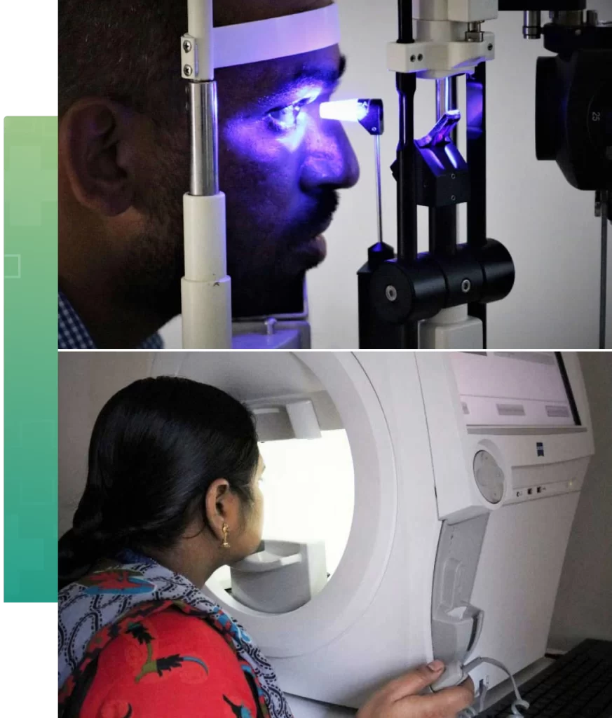
A comprehensive glaucoma evaluation includes the following tests which help in finding out the presence of glaucoma and also its progression in the subsequent follow-ups.
Tonometry is a diagnostic test that measures the fluid pressure, known as intraocular pressure (IOP), inside your eye. The tonometry test is important as it can help your doctor evaluate whether or not you may be at risk of glaucoma. In a diagnosed patient of glaucoma this is important for understanding the risk of progression and if the treatment is effective.
Gonioscopy is a painless eye examination of the front portion of your eye (anterior chamber), to examine whether the area where fluid drains out of your eye (drainage angle) is open or closed. This test is important as it helps your doctor to diagnose and tailor the kind of treatment most effective for your glaucoma.
The Visual Field test (Perimetry) produces a map of your complete vision. This test will help your doctor measure your peripheral or side vision (which is affected first by glaucoma), through which they can diagnose and monitor glaucoma. The data from the test is used to determine the severity of your glaucoma, level of vision loss, damage to the visual pathways of the brain, and other optic nerve diseases.
This diagnostic procedure helps the doctor examine your optic nerve for glaucoma damage. Eye drops are used to dilate the pupil so that the doctor can see through your eye to examine the shape and color of the optic nerve.
Optical Coherence Tomography of the optic nerve head is a noninvasive imaging technique widely used in the diagnosis and management of glaucoma. It provides high-resolution cross-sectional images of the optic nerve head, retinal nerve fiber layer (RNFL), and ganglion cell complex, allowing early detection of glaucomatous damage before visual field defects become apparent. OCT is indicated in suspected glaucoma, ocular hypertension, glaucoma suspects with suspicious optic discs, and for monitoring progression in established glaucoma cases. The major advantages of OCT include rapid image acquisition, objective quantitative analysis, reproducibility, and the ability to detect structural changes early. It is also useful for serial follow-up and progression analysis. However, OCT findings should always be correlated with clinical examination and visual field testing for accurate diagnosis and management of glaucoma.
Apart from the above routine glaucoma tests, your doctor may advice further investigations based on the type and severity of your glaucoma.
Narayana Nethralaya employs the most advanced diagnostic tools and investigational facilities needed in the evaluation and diagnosis of several common and rare kinds of glaucoma.
After methodological assessment of patients, doctors discuss the best available treatment options ranging from topical eye drops, oral medications or interventional procedures like lasers or surgeries. Patients are encouraged to ask questions and doctors go the extra mile to answer all their apprehensions.
All these new investigational modalities help us to detect the disease and progression early which aides us in better management of the patient.
If you have a family history of Glaucoma, are diabetic, have been on any form of steroids, are experiencing intense throbbing pain in the eye along with redness or notice decrease in vision, it is a good time to schedule an appointment with an ophthalmologist to get your eyes checked for glaucoma. The doctors at Narayana Nethralaya will evaluate the health of your eyes and assess your glaucoma. Your initial consultation will approximately take 2 hours if you do not require cross-consultation and up to 4 hours if you require cross-consultation. During your consultation, our doctors and counsellors will determine the best course of action for your visual needs, go over the risks and benefits of glaucoma treatment, and help you choose the best procedure that is suitable for preserving your visual needs. We suggest you bring a family member or friend with you to help you with your decision-making.

Damage caused by Glaucoma cannot be reversed, but early treatment and regular check-ups can help slow or prevent vision loss especially in the initial stage. Glaucoma is treated by lowering your eye pressure (IOP-Intraocular Pressure). Depending on the stage of your disease, your options may be eye drops, oral medications, laser treatment and surgery or a combination of any of these.
Your doctor will start by prescribing eye drops that will help decrease your eye pressure. This is either by improving fluid drainage from the eye or by decreasing the amount of fluid your eye makes. Depending on how low your eye pressure needs to be, more than one eye drop may be prescribed.
Some of these drops can cause mild irritation in the eyes, burning, redness, or pigmentation. Rarely, eye drops can also cause other side effects by getting absorbed systemically. Discuss with your doctor if you are not comfortable with your drops.
Laser peripheral iridotomy is the standard treatment in closed-angle glaucoma and eyes at risk for this condition. In this procedure, the laser beam creates a hole in the colored part of the eye (iris), leading to an alternative pathway for the outflow of fluid.
Minimally Invasive Glaucoma Surgery (MIGS) represents the cutting-edge approach in glaucoma treatment, emphasizing reduced invasiveness. During this procedure, a tiny tube or stent (known as the iStent) is delicately inserted into the eye. Alternatively, excess tissue can be removed using specialized techniques like the Bent Ab-Interno Needle Goniectomy (BANG) or the Kahook Dual Blade, allowing the fluid inside the eye to flow out smoothly. This advanced method effectively reduces intraocular pressure, aiding in glaucoma management.
MIGS is often recommended for individuals with mild to moderate glaucoma, aiming to provide significant relief while minimizing the disruption caused by traditional surgical methods.
A small hole is made in the wall of the eye which is guarded by a flap that acts as a trap door. The fluid is shunted from inside the eye in a controlled fashion, which forms a small ‘bleb’ underneath the upper eyelid. It is a surgical procedure to lower the eye pressure when medical therapy or laser treatment has failed.
Glaucoma valve surgery is used to manage moderate to severe or treatment-resistant glaucoma by implanting a drainage device that helps lower intraocular pressure.
Common implants include the Ahmed Glaucoma Valve (AGV), Ahmed Clear Path (ACP) and the Aurolab Aqueous Drainage Implant (AADI). These devices create an alternative pathway for the aqueous fluid to drain from the eye into a reservoir (bleb) beneath the conjunctiva.
Trans Scleral Cyclophotocoagulation (TSCPC) is typically reserved for severe or refractory cases of glaucoma. It involves destroying a part of the ciliary body – the structure that produces aqueous humor to lower IOP.
Cyclo G6 Laser procedure works on the same principle as TSCPC but the micro pulse mode delivers short, repetitive bursts of laser allowing issue cooling between pulses which minimises the damage and preserves the surrounding tissues. This makes it more safe and more repeatable.
In today’s world, people are quite aware about how lifestyle choices affect our overall health, and the same goes for glaucoma. Lifestyle choices can, to some extent, influence eye pressure and affect the risk of developing glaucoma, but there aren’t enough proven studies about the same in glaucoma patients.
Research indicates that regular exercise and an active lifestyle can help reduce their risk of glaucoma. Activities which raise your pulse by 20 to 25, like walking briskly, can help reduce IOP. Aerobics is known to cause a temporary decrease in the intraocular pressure but it has not been tested in glaucoma patients. Swimming reduces vulnerability of the optic nerve to an increase in the intraocular pressure.


A diet rich in green leafy vegetables is supposed to lower eye pressure.
Food items like shellfish, tuna, nuts, and seeds are rich in DHA (docosahexaenoic acid) and are helpful in preserving a healthy retina.
A diet rich in spinach, broccoli, and sprouts, which contain lutein and zeaxanthin, also helps avoid oxidative damage to the optic nerve.
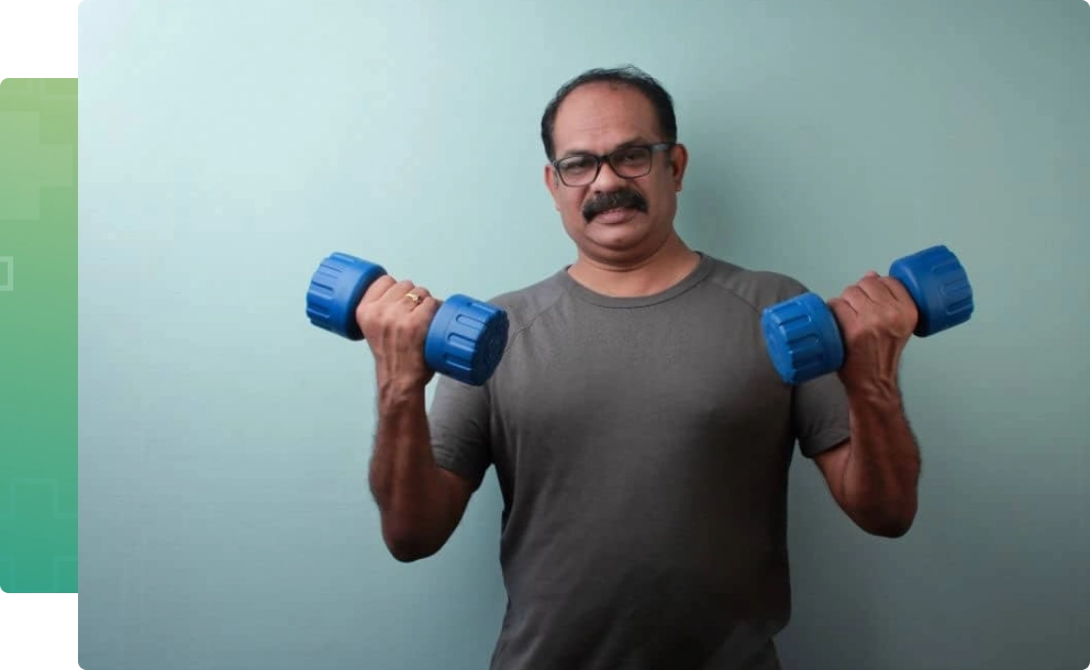
Lifting of heavy weights is known to cause a temporary increase in the intraocular pressure. Hence, patients who already suffer from glaucoma are advised to avoid lifting very heavy weights. Also some of the yoga asanas, particularly those with the head – down position (sheershasana) have been proven to cause an increase in the eye pressure.
Yoga headstands are generally not recommended for people with glaucoma due to the potential for increased intraocular pressure (IOP).

Foods which are rich in trans-fats, the kind found in deep fried food, prevent the optimal functioning of omega 3- fatty acids and increases eye pressure.
Hydrotherapy in the context of drinking water can benefit overall health, but in glaucoma patients, it’s important to avoid consuming large quantities of water in a short time. Rapid intake (over 1 liter in less than 15–20 minutes) can temporarily raise intraocular pressure, which may worsen glaucoma.
It’s best for glaucoma patients to sip water slowly throughout the day rather than drinking large amounts at once.

Caffeine, alcohol and tobacco are known to have a negative effect on eye pressure. Caffeine, when taken in excessive quantities has been proven to increase the risk of glaucoma. Daily consumption of alcohol and smoking cigarettes are detrimental to the optic nerve.
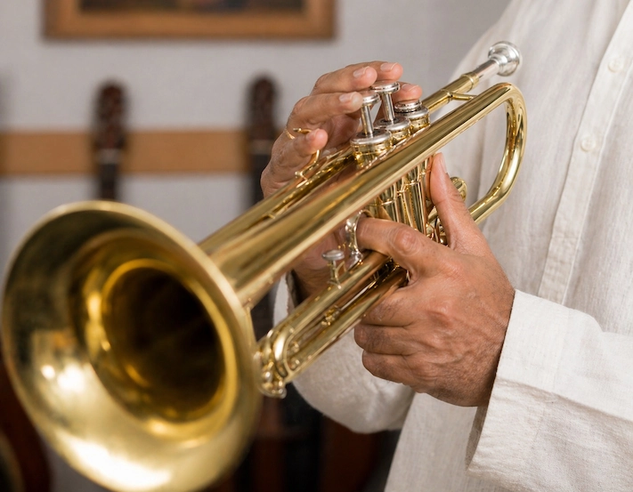
The trumpet and the saxophone are known to cause an increase in the eye pressure.
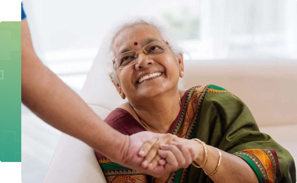
Glaucoma affects the quality of life of millions of people. Some forms of glaucoma may present with sudden onset of symptoms like eye pain, coloured halos, redness, and blurring of vision whereas in most cases, patients may not experience any symptoms in the early stages. It is only at the advanced stage of the disease that the patient begins to perceive the visual disturbances associated with glaucoma. Ocular discomfort, psychological factors, economic factors, and social constraints contribute to the burden of the disease.

A better understanding of your treatment and medications can make it easier to live with glaucoma and easier for your doctor to control your disease. Early diagnosis, regular follow ups and therefore early intervention, is crucial in delaying the progression.
If you have undergone any surgery or laser treatment for glaucoma, it is important to understand that these procedures are performed to reduce the intraocular pressure and are an attempt to restore the remaining vision.
Follow the instructions of your doctor and attend follow-up examinations as advised.
Follow the technique properly while instilling the medication into the eye so that the desired effect of the medication is attained.
Regular usage of medications:
– Keeping alarms on your mobile phone will remind you to instil your medications on time.
– Making a simple drug chart in a pocket diary, as shown in the figure, so that it can be noted and shown to your doctor as well.
If you have any difficulty in instilling the medication or if you have any discomfort after using it, let your doctor know about it.
You may not appreciate a change in vision, but never stop the medications without consulting your doctor; this can lead to further vision compromise.
There is no single value to be considered as normal eye pressure. On an average your eye pressure should be within 10-22mmHg but it also depends on various other factors like gender, age, ethnicity, hereditary, corneal thickness to name a few.
Every individual above the age of 40 years must have an annual ophthalmic evaluation, however glaucoma screening is must if there is a family history of glaucoma, systemic disease such as diabetes/hypertension, high myopia, history of trauma to the eye, long term steroid use.
The frequency of follow up is usually decided by your doctor, based on the severity of the disease. It is important to ensure regular follow ups to prevent worsening of the disease.
No,the existing nerve damage is not reversible. However, timely intervention and regular follow ups can prevent the progression of the disease.
Most people with glaucoma can still drive. Your ability to drive will depend on how much field of vision has been lost. Your doctor will perform regular visual field tests to evaluate the same.
You must continue routine timely instillation of medications until the earliest time you can visit your doctor.
To minimize systemic absorption, close your eyes for one/ two minutes after putting the eye drops. You may then press lightly at the corner of your eye near your nose to close the tear duct for one/ two minutes. Wipe off excess eye drops from your eyelid. If you have more than one eye drop, space them at 5 minutes interval between each drop.
It is usually rare. However,due to systemic absorption some individuals may experience dizziness, decreased heart rate, excacerbation of asthma, impotence, tingling sensation of extremities, fatigue, metallic taste, frequent urination, dry mouth etc. depending on the medication you are using. Always consult your doctor if you experience any new systemic effects after starting any medication.
Angle is the space between the clear part (cornea) of the eye and coloured part (iris) of the eye through which the aqueous flows out of the eye.
In closed angle glaucoma the angle becomes narrow or closed causing either sudden increase in eye pressure or a gradual increase which could lead to optic nerve damage and possible vision loss.
It is done as an OPD procedure.
There could be temporary blurring of vision, redness of the eye or increases sensitivity to light. These usually subside within a day. Anti-inflammatory drops are prescribed after the procedure and the eye pressure is assessed after 1 week.
Not always. In case of an acute attack of angle closure, the patient experiences blurred vision due to collection of fluid in the clear part of the eye( cornea). The peripheral iridotomy helps relieve the blockage of fluid outflow and clears the cornea. This restores the vision to pre attack status. However the procedure is meant to preserve the existing vision and prevent glaucoma progression.
In majority of cases, the procedure carries little to no risks. The documented risks are closure of irodotomy requiring re-treatment, excessive bleeding (especially in patients on blood thinners), progression of cataract, rarely retinal and choroidal detachments. Your doctor will discuss your risk profile with you and take necessary precautions.
Although it is unusual, serious complications such as infection, bleeding in the eye, loss of vision and eye pressure that is too low or high can occur. Therefore, regular follow ups to monitor the eye pressure, surgical wound and bleb along with disease progression is important even after the surgery.
Most patients do not require medications to control the eye pressure post trabeculectomy. Some patients continue to progress despite surgical intervention. In such patients if your doctor feels the eye pressure is not optimal or if there is a progression you may be advised to continue using the eyedrops.
Usually it is a safe procedure to control the eye pressure, but certain complications like infection, tube extrusion, corneal decompensation, implant failure may occur. These complications can occur any number of years post surgery. Hence
regular follow up as advised by your doctor is important to optimize your results.
It is a relatively safe procedure, complications like inflammatory reaction, pain, red eye, conjunctival scarring, extremely low eye pressure and further vision loss may occur in rare cases.
POAG
A 46 year old gentleman came to NN for glaucoma screening as his father had been diagnosed and treated for glaucoma at NN. After detailed ocular examination and glaucoma investigations, he was noted to have features suggestive of open angle glaucoma in both eyes. Topical anti glaucoma medications were started. As a result of timely intervention, on subsequent visits his eye pressure, vision and other glaucoma investigations remained stable. He has been advised to follow up regularly.
Congenital Glaucoma
A 6months old male child was referred to NN with a history of whitish discoloration, watering and enlargement of both eyes. The child was evaluated in the glaucoma clinic and diagnosed with congenital glaucoma. He underwent evaluation under general anesthesia and glaucoma surgery to control his eye pressure. He is 7 years old now and his eyes pressures and vision are stable. He continues to be on regular follow up. Early diagnosis and management has helped in preventing blindness in this child.
PACG
A 52-year-old woman presented to NN glaucoma services with complaints of severe pain and redness in the right eye associated with headache, vomiting and drop in vision. After a thorough clinical ocular examination her eye pressure was noted to be high and she was diagnosed with acute angle closure attack in the right eye. She was then advised laser iridotomy in both eyes along with topical and oral anti glaucoma medications.
When she was reevaluated the next day her eye pressure in the right eye was optimal and she was symptomatically better on subsequent visit her eye pressure and vision remained normal and she was advised regular follow ups.
At Narayana Nethralaya, “Quality of Care” and “Patient Safety” is our priority. Concern for our patient’s well being is at the core of what we do, and what drives us. Five units of Narayana Nethralaya are NABH Accredited – the highest national recognition for quality in patient care and safety. Our Glaucoma team help patients make an informed treatment choice on the type of treatment and surgery that is best suited for their lifestyle. We have an exclusive counseling team to address any doubts or questions that people may have about Glaucoma treatment options, procedures, preoperative testing and post surgery recovery.